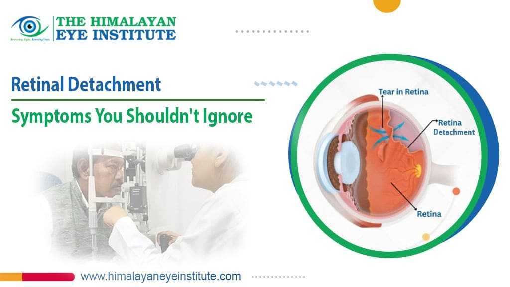


Retinal detachment is a medical emergency where the retina pulls away from the back of the eye - causing sudden vision changes that, if left untreated for even a few hours, can lead to permanent vision loss in the affected eye.
Most people who lose vision to retinal detachment didn't see it coming. Not because there were no warning signs - there almost always are - but because the signs are easy to dismiss. A few new floaters. A flash of light out of the corner of your eye. A shadow at the edge of your vision.
Each one on its own seems harmless. Together, they're your eyes telling you something is wrong.
This is the condition where waiting costs you. Not days - sometimes hours.
What Is the Retina and Why Does Detachment Matter?
The retina is the thin layer of light-sensitive tissue lining the back of your eye - it converts what you see into signals sent to your brain. When it detaches, those signals stop, and vision in that area is lost.
Think of the retina like the film inside a camera. The lens focuses the image, but without the film - or with film that's come loose - no picture forms. The retina works the same way. It's what actually processes everything you see.
When it begins to detach, the part of your vision controlled by that area starts to go. The process can happen slowly over days or suddenly over hours, depending on the type of detachment. Either way, it doesn't reverse on its own. Without surgery, the detachment typically progresses until vision in that eye is gone.
The good news - and this is worth holding onto - is that retinal detachment surgery succeeds in approximately 9 out of 10 cases when treatment begins promptly. The outcome is directly tied to how quickly you act.
Early Warning Signs of Retinal Detachment
The most common early warning signs of retinal detachment are a sudden increase in floaters, flashes of light in peripheral vision, and a dark shadow or curtain appearing at the edge of your visual field.
These symptoms often appear without any pain - which is part of why people underestimate them. A painless eye rarely feels urgent.
Here's what to watch for:
Sudden floatersare the small specks, threads, or cobweb-like shapes that drift across your vision. Most people have a few and they're harmless. But a sudden increase in floaters - especially a shower of new ones appearing at once - is a red flag. Floaters are reported in 70 to 90% of retinal detachment cases.
Flashes of light Brief, lightning-like flashes in your peripheral vision - often described as seeing sparks or a camera flash in the corner of your eye. They can happen in the dark, which is when people notice them most. These flashes happen when the vitreous gel pulls on the retina.
A shadow or dark curtainThis is the symptom that most clearly signals active detachment. A dark shape - described by patients as a curtain, shadow, or grey cloud - creeping in from the side, top, or bottom of your vision. It may start small and grow over hours.
Blurred or distorted visionCentral vision can become blurry or wavy, as though looking through frosted glass or water.
Grey or dark area in your visual fieldA portion of your vision simply goes dark - not from the outside in like a curtain, but a fixed dark patch that stays there.
One Thing Every Person Should Know About These Symptoms
If you're seeing new floaters and flashes together - especially alongside any shadow in your vision - don't wait until morning. Don't wait until your regular appointment.
Go to an eye doctor the same day.
Retinal detachment will not heal by itself and waiting will delay further progression of the condition. If the condition progresses to the involvement of the macula - the central area which is responsible for clear and sharp vision, it will lead to a poorer prognosis if treated at that stage.
The gap between an early diagnosis and a late diagnosis may be the gap between restoring sight fully and restoring sight partially.
Does Retinal Detachment Hurt?
No - retinal detachment is typically painless. The absence of pain is one of the reasons people delay seeking help, mistaking a serious eye emergency for something minor.
This is one of the most important things to understand about this condition.
Most medical emergencies hurt. A heart attack, a broken bone, appendicitis - pain is what sends people to the hospital. Retinal detachment doesn't work that way. Your eye can be in the process of losing its vision and feel perfectly comfortable.
If you're waiting for pain to tell you something is wrong, you may already be too late.
Types of Retinal Detachment
There are three types of retinal detachment - rhegmatogenous, tractional, and exudative - each with a different cause and slightly different presentation.
|
Type |
What Causes It |
Who's Most at Risk |
|
Rhegmatogenous |
A tear or hole in the retina lets fluid underneath, lifting it away |
Most common type - affects people with high myopia, aging vitreous, or after eye injury |
|
Tractional |
Scar tissue on the retina pulls it away from the back of the eye |
People with diabetic retinopathy, previous eye surgery |
|
Exudative |
Fluid accumulates beneath the retina without a tear - from inflammation or leaking blood vessels |
People with inflammatory eye conditions, high blood pressure, or tumors |
Rhegmatogenous is by far the most common - it accounts for the majority of cases and is the type most associated with the classic warning symptoms of floaters, flashes, and a shadow in vision.
Who Is at Higher Risk?
Retinal detachment can affect any person, but there are specific factors that predispose a person to retinal detachment, including high myopia, age, history of eye surgery, eye injury, diabetes, and familial predisposition to retinal detachment.
You should be especially vigilant about the warning signs if you:
- Are significantly short-sighted (myopia above -6 diopters)
- Are over 50 - the vitreous gel naturally shrinks and can pull away from the retina with age
- Have had a previous retinal detachment in either eye
- Have a family history of retinal detachment
- Have had cataract surgery or other eye procedures
- Have diabetes - diabetic retinopathy is a leading cause of tractional detachment
- Have experienced a direct eye injury or trauma
- Have posterior vitreous detachment (PVD) - a common age-related change that can lead to retinal tears
Being in a higher-risk group doesn't mean retinal detachment is inevitable. It means regular retinal examinations are not optional.
How Is Retinal Detachment Diagnosed?
Retinal detachment is diagnosed by an ophthalmologist using a dilated eye examination - the retina cannot be fully evaluated without dilation, which is why regular dilated eye exams matter even when your vision seems fine.
The examination uses a bright light and lenses to look at the retina directly. In some cases, an ultrasound scan of the eye is used if the retina isn't visible due to bleeding inside the eye.
The entire examination is quick. It's the most important thing you can do if you have even a slight suspicion of retinal detachment. A general optometrist can perform an initial check, but a retina specialist at a dedicated retina hospital in Siliguri is who you want for diagnosis and treatment.
Treatment Options for Retinal Detachment
Retinal detachment always requires surgical treatment - it will not heal on its own, and the type of surgery depends on the severity, location, and type of detachment.
If caught very early as a retinal tear before detachment has occurred, laser treatment or cryopexy (freezing) can seal the tear and prevent detachment from progressing.
Once detachment has occurred, the main surgical options are:
Pneumatic RetinopexyA gas bubble is injected into the eye to push the retina back into position. Laser or cryopexy seals the tear. The patient must hold a specific head position for several days while the bubble does its work. Used for certain types of detachment with tears in the upper part of the retina.
Scleral BucklingA silicone band is placed around the outside of the eye to gently push the eye wall inward, relieving the traction on the retina. Often combined with draining the fluid beneath the retina. One of the oldest and most reliable surgical treatments for retinal detachment.
Vitrectomy Vitrectomy is when the gel is removed from the back of the eye and replaced with either a gas bubble or silicone oil to maintain the position of the retina while it heals. This is the most adaptable form of treatment and is used in complicated detachments and traction cases.
|
Treatment |
When It's Used |
Recovery |
|
Laser / Cryopexy |
Retinal tear - before full detachment |
Minimal, outpatient |
|
Pneumatic Retinopexy |
Specific upper-retina detachments |
Days of strict positioning |
|
Scleral Buckling |
Rhegmatogenous detachment |
Several weeks |
|
Vitrectomy |
Complex or tractional detachment |
Several weeks, gas/oil in eye |
Retinal detachment surgery is usually successful in about 9 out of 10 patients. The success of vision restoration will depend on whether the macula was damaged prior to the surgery, the duration of the condition, and the general health of the retina. This is why speed matters more with this condition than almost any other.
What Happens If Retinal Detachment Is Left Untreated?
Without treatment, retinal detachment progresses to complete vision loss in the affected eye - the retina deteriorates over time once separated from its blood supply, and this damage becomes permanent.
The retina needs a constant supply of oxygen and nutrients from the tissue it sits against. Once it detaches, it's cut off. The longer it stays detached, the more the retinal cells die.
In the early stages, some vision can usually be recovered. Once the central macula is involved - or once the detachment has been present for weeks - the vision that's been lost may not come back even after successful surgery.
There is no medication, no eye drop, and no home remedy that reverses retinal detachment. Surgery is the only option.
Frequently Asked Questions
Can retinal detachment heal on its own?
No. Retinal detachment cannot heal without surgery. If you have signs, then you should seek treatment immediately to prevent loss of vision.
Is retinal detachment the same as a retinal tear?
Not at all – a retinal tear is an injury to the retina, which, left unattended, could let fluid leak underneath the retina and result in its detachment. The earlier this tear is detected, the sooner treatment by laser surgery or cryopexy can take place.
How quickly does retinal detachment progress?
It all depends. Detachments can take days to occur; some can occur within hours. It’s impossible to predict how quickly it will happen in any one case, and that’s why it’s recommended to be seen that day when symptoms arise.
Can retinal detachment affect both eyes?
It commonly affects one eye at a time, but individuals that have suffered from retinal detachment in one eye run a higher risk of getting detached in the other. Follow-ups of both eyes are very important after experiencing retinal detachment.
What does vision look like with retinal detachment?
Patients describe seeing a shadow or curtain falling over their field of vision, sudden appearance of floaters, flashes of light, and blackouts in certain parts of their vision. Some patients describe seeing things through a veil within their field of vision.
Can short-sighted people get retinal detachment?
Yes - and they're at significantly higher risk. High myopia stretches and thins the retina, making it more prone to tears and detachment. Regular retinal examinations are strongly recommended for anyone with significant short-sightedness.
Retina Treatment in Siliguri - Himalayan Eye Institute
Retinal detachment is one of those conditions where what you do in the first few hours genuinely determines the outcome. Not the first few days. The first few hours.
If you or someone you know has sudden floaters, flashes of light, or a shadow appearing in their vision - don't wait to see if it passes. It won't pass. It will progress.
The Himalayan Eye Institute is North Bengal's most trusted eye hospital in Siliguri, with a dedicated retina unit staffed by experienced vitreoretinal surgeons. Whether you need urgent evaluation for suspected retinal detachment or advanced retina treatment in Siliguri, our team is equipped to diagnose and treat at the level your vision deserves.
As a NABH-certified eye doctor Siliguri center with a 4.9 rating across 9,000+ reviews, we've seen what a difference early treatment makes - and we've seen what happens when people wait too long.
Don't wait.
Address: Jhankar More, Burdwan Road, Siliguri – 734005
Appointments: +91 74077 40723 | +91 89007 99992
Online Booking: appointment.himalayaneyeinstitute.com